I Have a Blue Nodule on My Arm
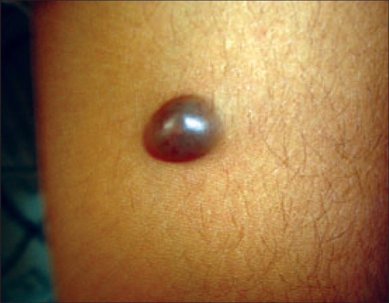
A 21-year-old male presented with an asymptomatic, slow-growing swelling on the left arm of 4 years duration. He gave history of expression of black-colored fluid and, sometimes, blood when punctured. On examination, solitary, sessile, globular, well-circumscribed bluish nodule, measuring 2 cm in diameter, was present on the lower part of his left arm [Figure - 1]. The surface of the nodule was smooth, with visible superficial vessels. On palpation, the nodule was freely mobile, nontender and soft in consistency, with firm base. An excisional biopsy showed a single cyst (3 mm) containing black-colored material. Histopathological features are shown in [Figure - 2],[Figure - 3].
Diagnosis: Nodular hidradenoma
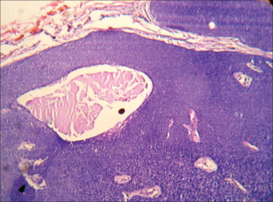
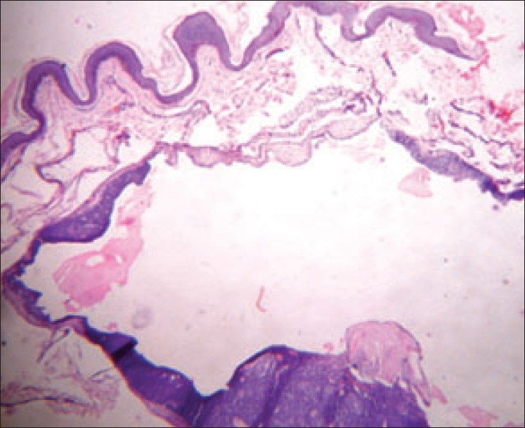
Histopathology showed well-circumscribed tumor with solid and cystic areas [Figure - 2], arranged in lobules separated by thin fibrovascular septae [Figure - 3]. Two types of tumor cells were seen. One was oval-to-polyhedral with round-to-oval basophilic nucleus and moderate amount of eosinophilic cytoplasm. The other was round-to-oval with small hyperchromatic nucleus, and moderate amount of pale cytoplasm was also seen within the tumor. There were cystic spaces of varying sizes containing mucinous material lined by single-to-multilayered cuboidal epithelium. At some areas, degenerating tumor cells secreting their contents into the cystic spaces were noted.
Discussion
Nodular hidradenomas (NHs) are primitive adnexal tumors that show differentiation towards eccrine and apocrine apparatus. [1] Clinically, NH presents as slow-growing, firm, solitary, bluish-red mobile nodule, mainly situated over scalp, face, trunk and occasionally on the extremities. [2] Rarely, it may be cystic, pedunculated or ulcerated. [3] The size of the nodule varies from 5 to 30 mm in diameter. [1] However, a size up to 10 cm has been reported. [3] The overlying skin is stretched and atrophic with visible surface vasculature. [4],[5],[6] NH is usually seen in adults with an average age of 37.2 years and a female-to-male ratio of 1.7:1. [7] Though usually seen between the fourth and eighth decade of the life, cases have been reported in young children.
Since there are no specific features to diagnose NH clinically, the histopathological study is essential for the correct diagnosis. Microscopically, NH is a circumscribed noncapsulated dermal tumor, sometimes extending into the subcutaneous tissue, with solid areas, cystic spaces and tubular lumina. The solid area is characterized by three types of cells: polyhedral cells with round nucleus and basophilic cytoplasm, similar to cuboidal ductal cells; fusiform cells with elongated nucleus located at the periphery of the tumor, representing myoepithelial cells; and round cells with small dark nucleus and clear cytoplasm, representing secretory coil cells. Occasionally there may be foci of squamoid differentiation with horn pearls, representing intraepidermal ductal cells. [8] Similarly, tubular lumina are also lined by four types of cells representing cells of different parts of the sweat gland. [3] Often there are cystic spaces of considerable size bordered by tumor cells. Formation of these cystic spaces is attributed to the degeneration of tumor cells. Sometimes, tumor nodules show areas of eosinophilic hyalinized stroma. [8] Other cellular variants include oncocytic, epidermoid and pigmented variants with melanocytes and melanin pigment in the cells and macrophages. [9]
Multiple foci of growth, atypical mitoses, zonal necrosis and invasion of vascular and perineural sheath indicate the possibility of hidradenocarcinoma. [1] Histologically, tricholemmoma also shows clear cells. However, tricholemmoma shows peripheral palisading of tumor cells with the absence of cystic spaces and tubular structures. [8]
On fine needle aspiration cytology (FNAC), NH is usually misdiagnosed as breast carcinoma when it presents as breast lump with ulceration [10] and as cutaneous metastasis of conventional-type renal cell carcinoma. [11] Since such cases are usually referred to FNAC, the knowledge of cytological features of NH can aid in the proper diagnosis of the condition. On FNAC, NH is diagnosed by the presence of many clear cells amidst polygonal tumor cells. [10] NH may also arise from nevus sebaceous as a result of dysembryoplasia [12] and from mammary duct as subareolar nodule with nipple discharge. [13]
NH is a benign tumor but malignant transformation with metastasis has been reported. [14] Local recurrences can occur if the tumor is incompletely excised. [9] Though mitoses do not necessarily indicate the malignancy, their presence is associated with increased local recurrences and subsequent malignant transformation. [9] Surgical excision is the recommended treatment. [14]
Source: https://ijdvl.com/a-bluish-nodule-on-the-arm/
0 Response to "I Have a Blue Nodule on My Arm"
Post a Comment